← All courses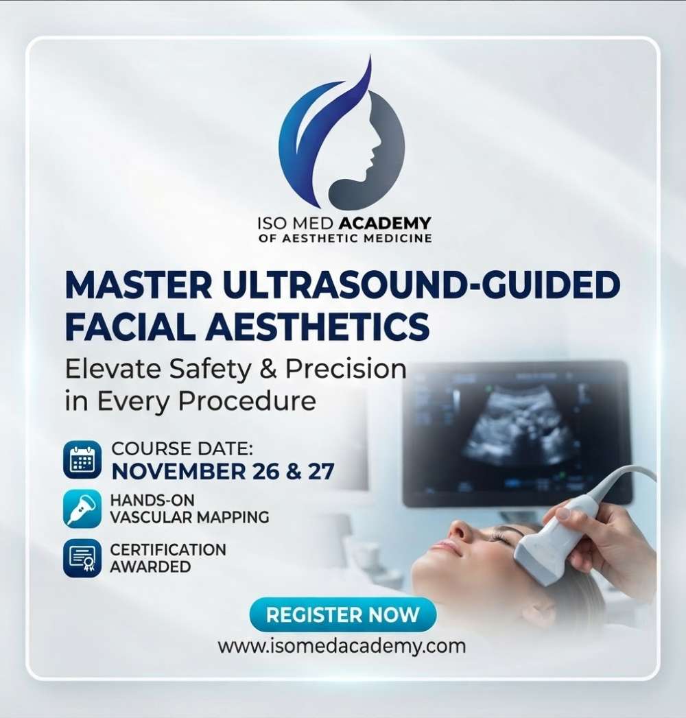
ISO MED ACADEMY
Designing an Ultrasound-Guided Facial Aesthetics Course
Integrating ultrasound into facial aesthetics elevates practice safety by enabling real-time vascular mapping, filler localization, and complication management. Here is a structured, comprehensive curriculum outline tailored for clinical professionals.
Course topics — objectives of the course
- Target Audience: Physicians, dentists, and advanced aesthetic practitioners performing facial injectables.
- Core Goal: Master high-frequency ultrasound imaging for facial anatomy, filler identification, and complication avoidance (e.g., vascular occlusion).
- Format Recommendation: 1 to 2-day hybrid model combining didactic lectures, live scanning demonstrations, and hands-on transducer work.
Course program: Module 1: Physics, Instrumentation & Ultrasound Basics
- Principles of Ultrasound: Piezoelectric effect, frequency selection (\bm{10\text{--}22\text{ MHz}} linear transducers), and image optimization (gain, depth, focus).
- Machine Settings & Artifacts: Understanding color Doppler, power Doppler, and acoustic shadowing.
- Probe Handling: Ergonomics, transducer stabilization, and appropriate gel application techniques.
Course program: Module 2: Applied Facial Anatomy & Sonographic Mapping
- Upper Face: Supraorbital/supratrochlear arteries, frontal branch of the temporal artery, and pericranial layers.
- Mid Face: Infraorbital artery, zygomatico-facial artery, transverse facial artery, and SMAS/deep fat compartments.
- Lower Face & Jawline: Facial artery trajectory along the mandibular border, labial arteries, and mental foramina.
- Danger Zones: Identifying high-risk anatomical corridors versus safe fascial planes in real time.
Course program: Module 3: Identifying Product Types & Complications
- Product Sonographic Appearance: Differentiating hyaluronic acid (HA), calcium hydroxylapatite, PLLA, and permanent fillers under ultrasound.
- Complication Assessment: Detecting acute vascular compromise, persistent edema, late-onset nodules, and biofilm formation.
- Dissolution Guidance: Real-time ultrasound guidance for precision hyaluronidase placement in vascular occlusion management.
Course program: Module 4: Hands-on Practical Workshop
- Live Demonstration: Instructor-led live scanning on models mapping major facial arteries.
- Small-Group Scanning: Participant-to-participant or model-based scanning sessions focusing on probe placement over standard injection zones.
- Dynamic Maneuvers: Assessing tissue movement during animation and identifying filler integration versus displacement.
Course program: Recommended Teaching Materials
- Pre-Course Reading: Recommended articles on facial sonography and vascular anatomy landmarks.
- Assessment: Short pre- and post-course knowledge quizzes to evaluate competency gains.
- Certificate of Completion: Awarded upon successful attendance and practical skill demonstration.